Technology edge
AB ultrasound scanner (Biomedix echovue, India (Japan collaboration)
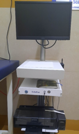
This is a new generation ultrasound that works on the principle of action of high frequency sound waves converted into electrical signals with high quality built in imaging system software, convenient and portable incorporating smart tools that increase reproducibility. Special features are dynamic recording and video capturing dual mode of scanning for the purpose of accurate intraocular lens power calculation in cataract operations and to diagnose early, the retinal causes for blindness in childhood and adults for intraocular tumours and retinal detachments.
The most advantage is that retina is visualised effectively in the presence of corneal scars and opacities, dense cataracts and lid problems where retinal visualization is not possible with HD imaging and dynamic recordings. It provides excellent patients care with superb imaging needs, of its innovative design providing consistence performance. Real time snapshots and video capture files are available for assessing the patients for the progression and treatment effectively. It helps to annotate patients’ images with clinical findings and communicator readily.
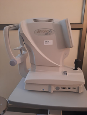
Auto-Kerato refractometer (TOPCON, Version RM 800 and KR 800, Japan)
This advanced device (TOPCON, version RM 800 and KR 800, Japan) incorporates two soft wares that measure not only refractive state of the eyes but also the corneal curvature, thus facilitating the quick diagnosis of short sightedness, long sightedness, astigmatism, presbyopia and keratoconus. When infra-red light falls on the retina, reflected rays of light is captured by inbuilt camera and the internal computer will calculate the values are the basic principle of this instrument. The greatest advantage of the machine is its simplicity of recording the data that can be computed for the research purpose.
Colposcopy

Cervical cancer is one of the major public health problem in India. India accounts for 15.2% of the total cervical cancer deaths in the world.Colposcopy is a screening and diagnosticprocedure to examine an illuminated, 40 times magnified view of the cervix as well as the vagina and vulva. The main goal of colposcopy is to prevent cervical cancer by detecting and treating precancerous lesions early. Screening for cervical cancer (either by Pap test and/or human papillomavirus testing) is an important part of staying healthy and avoiding cervical cancer. If the results of the screening test are abnormal, further testing is needed to confirm the result and determine the severity of the abnormality for which coploscopy is advised.A colposcopy is used to find abnormal lesion more accuratelyand thus helps in taking biopsy from that specific site for more accurate diagnosis. Treatment may be given in the same siting when indicated. Accurate diagnosis may also avoid the need for unnecessary hysterectomy.A colposcopy also looks for other health conditions, such as genital warts or polyps. Colposcopy is thus an invaluable method for detecting cancerous and precancerous lesion very early in the disease,saving many lives.
Continuous Renal Replacement Therapy (CRRT)
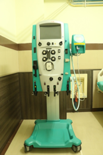
CRRT is used increasingly in Intensive care units across the world these days. The latest state of the art Gambro Prismaflex CRRT machine is being used for continuous renal replacement therapy in our hospital ICU. CRRT is a form of slow and continuous blood purification process used in patients with severe renal failure with hemodyamic instability ( low blood pressure) . As the name suggests, it is a form of continuous therapy performed for more than 24 hours and can be continued for several days. The major difference between the regular intermittenthemodialysis (IHD) and CRRT is the rate at which the water and wastes are removed from the blood. In regular hemodialysislarge amounts are removed in a short span of around 4 hours whereas in CRRT the removal takes place at a slow and steady pace because of which it is very useful in patients in low blood pressure. CRRT helps in better fluid and acid base management . This also helps in improving the hemodyamic stability of critically ill patients in an intensive care setting. This therapy is advantageous in patients requiring neurosurgical intervention as it helps in reducing the pressure within the brain.
iCARE tonometer (Icare ic100 Tonometer, Finland)
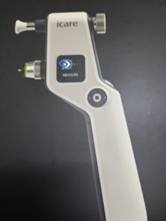
This is a newly invented non-contact method of measuring intraocular pressure that helps in very quick acquisition of readings facilitating the early diagnosis of glaucoma (increase in eye pressure that causes blindness due to pressure effect on the nerve of sight). Very thin sterilised disposable probe is used that makes a momentary impact on the cornea without contact which works on the principle of electromagnetic rebound technology and the most beneficial factor is that it is free from use of anaesthetic eye drops. It enables hygienic and effortless eye pressure measurements procedure that is barely noticed by the patients and is suitable for all age groups even works well for post-surgical patients.
Intra Operative Neurophysiological Monitoring Equipment
Modern neurosurgery has achieved great results in the field of brain and spine surgeries.This has been possible because of many technological advancements in the field of neurosurgery all around the world. To this end,Neurosurgery Department of JSS Medical College Hospital has not been laggingbehind.Apart from many modern equipments like Ultrasonic Aspirator and advanced Operative Microscope, this department has recently acquired,Neuromonitoring Equipment which is not available in any of the hospitals in and around Mysore city.
Neuromonitoring involves continuous evaluation of electrical activity of one or more neural pathways in an anesthetised patient.The goal of this monitoring is to detect even minor surgical or physiological insults to the brain and spinal cordduring the surgery so as to prevent any permanent damage which would have occurred without being aware of, by the operating surgeon.Neuromonitoring is an essential equipment for performing epilepsy [seizure disorder] surgery, brain aneurysm clipping,various skull base tumours like vestibular schwannoma which involve multiple cranial nerves and other intrinsic brain tumours involving important tracts from brain to the spinal cord .
Various diseases like tumours involving spinal cord substance,craniovertebral anomalies compressing the critical junction of brain with the spinal cord,complex spinal instrumentation surgeries,paediatric [child] developmental anomaly operations and peripheral nerve surgeries can all be performed with total accuracy and with the best results under the guidance of this neuromonitoring equipment.
Multi Para Monitoring
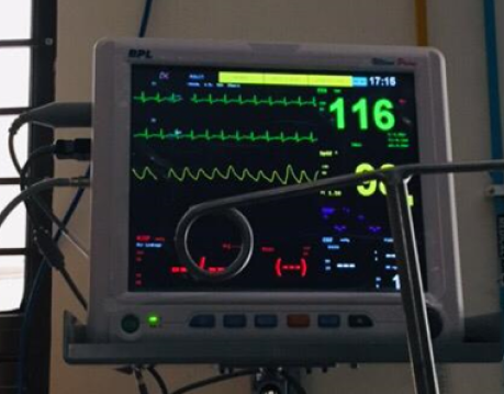

In medicine, monitoring is the observation of a disease, condition or one or several medical parameters over time. Monitoring of vital parameters can include several of the ones, and most commonly include blood pressure and heart rate, and preferably also pulse oximetry and respiratory rate. Multimodal monitors that simultaneously measure and display the relevant vital parameters are commonly integrated into the bedside monitors in critical care units, and the anesthetic machines in operating rooms. These allow for continuous monitoring of a patient, with medical staff being continuously informed of the changes in general condition of a patient.
Various parameter includes.
- Cardiac monitoring, which generally refers to continuous electrocardiography with assessment of the patients condition relative to their cardiac rhythm.
- Hemodynamic monitoring, which monitors the blood pressure and blood flow within the circulatory system. Blood pressure can be measured either invasively through an inserted blood pressure transducer assembly, or noninvasively with an inflatable blood pressure cuff.
- Respiratory monitoring, such as:
- Pulse oximetry which involves measurement of the saturated percentage of oxygen in the blood, referred to as SpO2, and measured by an infrared finger cuff
- Capnography, which involves CO2 measurements, referred to as EtCO2 or end-tidal carbon dioxide concentration. The respiratory rate monitored as such is called AWRR or airway respiratory rate)
- Respiratory rate monitoring through an ECG channel or via capnography
- Body temperature monitoring through an adhesive pad containing a thermoelectric transducer.
Multipara monitors are available in all Intensive Care Units in JSS Hospital and also in all the operation theatres ,in CT scan room, and post operative wards , and portable pulse oximeters in wards . so this enables us to effectively monitor the patient inside the hospital and improves the patient safety and helps to identify any deterioration of the patient early and manage the patient effectively.
Phototherapy Solution for Psoriasis and Vitiligo
Phototherapy is among the common therapeutic options in Dermatology for conditions like Vitiligo, Psoriasis, Eczema, Pruritus, etc. It is the use of ultraviolet radiation for therapeutic purposes. We have different modalities of phototherapy catering to different patient needs. Narrow band UVB phototherapy is mostly preferred. We have the whole body phototherapy chamber for patients having the skin lesions more generalized, involving skin trunk and limbs. We procured the new full body phototherapy unit from Daavlin ,USA.
It was inaugurated recently in the department of Dermatology at JSS Hospital , Mysore by Hospital Director Dr(Col) Dayanand , Principal –JSS Hospital Dr Basavanna Gowdappa in presence of Medical Spuritendent Dr M Guruswamy , DMS Dr Salah Ghori , HOD Dermatology Dr Verranna , Dr Jaydev Betkarur, Dr Shantamallappa and all the staffs Dermatology Dept.
The state of art equipment has 20 each of UVA and UVB light sources respectively. It can be used for PUVA & NBUVB treatment modalities. It can be used in all age groups. There are no side effects. The unit is fully computerized.
The facility is available at an affordable cost in department of Dermatology from 8:00am-7:30pm on weekdays and on sunday and Government holidays from 8:00am to 1:00pm
Apart from this, we do have the hand and foot unit for acral lesions and targeted phototherapy unit for localised delivery of phototherapy.

Zeiss Humphrey visual field analyser-3 (Model 840, Singapore)
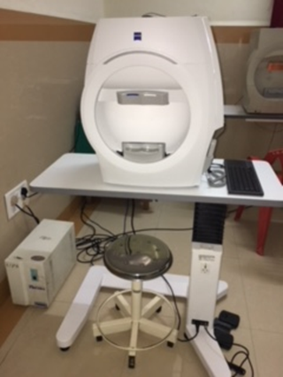
This is computerised automated visual fields analyser (SITA-Swedish interactive threshold algorithm) considered as Gold standard Test for early diagnosis of glaucoma that plots the boundaries of the field of vision which is affected and also in other neurological disorders leading onto blindness. This instruments aids in screening, monitoring of progressive visual loss and assisting in findings out new signs in the early stages of the disease process. The greatest advantages are reduced visual field testing time and simplified operation with a new smart touch interface in addition to the provision of new liquid trail lenses for correcting patients’ refractive error while undergoing test procedure.


